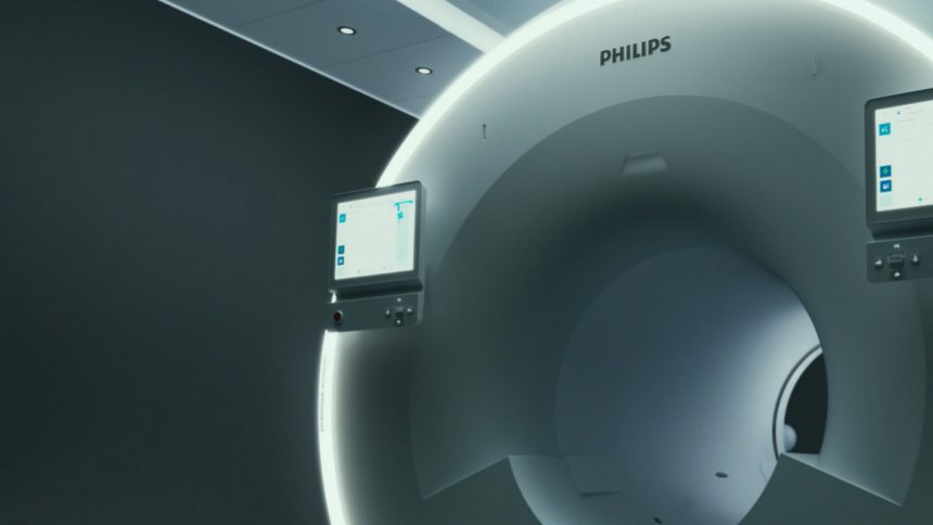
This fall, the Michigan State University Veterinary Medical Center will begin installation of a state-of-the-art MRI system that will allow the Hospital to take clearer-than-ever images of patients, providing a superior tool for veterinarians to diagnose medical issues accurately and to determine targeted treatment plans.

“The Philips MR 7700 is the first that Philips will have installed in a veterinary setting,” says Rebecca Linton, who manages the Hospital’s Radiology Service. “This puts us at the forefront of clinical imaging. MRI technology is constantly developing and advancing — with the new system’s features, we’ll be positioned to perform any type of imaging study that may be desired over the next 20 years.”
The system’s magnet has twice the field strength — going from 1.5T to 3T — of the Hospital’s previous MRI. (T, short for “Tesla,” is the unit that defines a magnet’s field strength.) The system also will use artificial intelligence and deep learning to refine both the scanning process and image quality. The machine is even capable of multi-nucleic imaging, a scanning innovation currently in its earliest stages.
The system, which will begin scanning patients of the MSU Veterinary Medical Center in early 2024, will be among the best available to veterinarians in the region. Patients will be referred by veterinarians near and far to take advantage of its capabilities.
Clear images, clear care pathways
Like in human medicine, MRI systems are used in veterinary medicine to diagnose pathologies such as tumors, strokes and injuries to bones and soft tissues. At the MSU Veterinary Medical Center, MRI is most often used to scan the brains and spines of neurology patients and to image tumors in oncology patients, but it has applications in other medical specialties.
“A 3T MRI is very powerful, with a high signal-to-noise ratio,” says Linton. “This means it gives great image quality, with more data points per voxel — the cube of tissue we’re imaging. This gives us the ability to scan even extremely small animals, like mice, to the largest subjects, like horses or wild cats.”
The new system’s power will likely expand the utility of MRI for medical specialties that historically have not utilized MRI as heavily.
“MRIs are the modality of choice to scan soft tissues, including body fat and water. When it comes to animals like horses, whose limbs are full of tough, difficult-to-image ligaments and tendons, a high signal-to-noise ratio provides better image quality,” says Linton. “This is going to open avenues for orthopedics, cardiology, abdominal and thoracic imaging — areas that the new MRI can image far better than before. This system will build out what we can do clinically for years to come.”
Improved image quality will give doctors more information that they can use to diagnose and prescribe treatments.
“Say for example we must scan a tiny cat patient in which some structures are sub-millimeter in size,” explains Jody Lawver, veterinary radiologist with the Hospital’s Diagnostic Imaging Service. “A higher signal-to-noise ratio provides the resolution needed to better see tiny structures, like nerves and cartilage; to identify whether they’re normal or abnormal. This additional diagnostic information helps us more fully characterize the condition and ultimately better guide therapeutics.”
With greater field strength and added technology, the new MRI will provide superior image quality 30–50% faster than before.
“Because we will be able to complete a standard brain study in, say, 20 minutes, we can add more MRI sequences to get more information in less time,” says Linton.
“These time savings can also be important for patient care,” Lawver adds. “Animal MRI requires general anesthesia, which can lead to a decrease in the patient’s body temperature and other problems like myopathy in horses. For heavy animals and patients that require surgery immediately after the MRI, it’s important to minimize MRI procedure time to ensure our patients remain warm, limiting complications. Our expert imaging, surgery, and anesthesia teams work closely together to balance imaging needs while maximizing patient care.”
Lawver points out how the new MRI capabilities will foster further innovation within veterinary medicine.
“Vet med is in the infancy state of some things that are mainstay in human medicine, so having extra time, superior image quality, and next-level technology from this machine allows us to achieve more clinical and research investigations. This allows us to collaborate with other leading universities with 3T magnets, which will undoubtedly move animal imaging forward.”
To learn more, visit the College of Veterinary Medicine website.